
 DNA Synthesis
 Vector Selection
 Molecular Biology
 Oligo Synthesis
 RNA Synthesis
 Variant Libraries
 Genome KO Library
 Oligo Pools
 Virus Packaging
 Gene Editing
 Protein Expression
 Antibody Services
 Peptide Services
 DNA Data Storage
 Standard Oligo
 Standard Genome KO Libraries
 Standard Genome Editing Plasmid
 ProXpress
 Protein Products
Anyone who has received an IV infusion knows how time-consuming it can be, especially when treatment requires repeated visits. What many people may not realize is that some large-molecule drugs, including antibody-based therapies, can be delivered subcutaneously rather than intravenously, reducing administration time from hours to just a few minutes.
So, what stands in the way?
One major barrier is the hyaluronic acid (HA)-rich extracellular matrix beneath the skin. And one enzyme has become particularly important in overcoming this barrier: recombinant human hyaluronidase PH20 (rHuPH20).
What Is rHuPH20?
Produced in CHO (Chinese hamster ovary) cells, rHuPH20 is a recombinant human enzyme consisting of 447 amino acids with a molecular weight of approximately 61 kDa. Its mechanism is remarkably straightforward. The subcutaneous tissue contains abundant hyaluronic acid, a highly hydrated, gel-like component of the extracellular matrix. This matrix helps maintain tissue structure but also limits the rapid movement of injected fluids.
rHuPH20 acts like a pair of molecular scissors. It temporarily breaks down HA into smaller fragments, reducing the density and viscosity of the local extracellular matrix. This creates a temporary window during which therapeutic molecules can spread more easily through the subcutaneous tissue and reach the blood and lymphatic vessels.
The result? Some drugs that once required hours of intravenous infusion can instead be administered through a subcutaneous injection in just a few minutes.
Even more importantly, the effect is local, temporary, and reversible. rHuPH20 acts primarily at the injection site, has a subcutaneous half-life of less than 30 minutes, and is rapidly cleared after entering systemic circulation. Because HA is also naturally turned over relatively quickly, the local tissue barrier can recover within approximately 24–48 hours.
This technology is already incorporated into several established therapies, including subcutaneous formulations of daratumumab, trastuzumab, and rituximab, as well as HYQVIA for long-term immunoglobulin replacement therapy. For many patients, this can mean spending minutes rather than hours in a clinic and, in some cases, administering treatment at home after appropriate training.
Understanding the “Opponent”
Hyaluronic Acid and the Extracellular Matrix
To understand why rHuPH20 is so useful, it helps to first understand its target: hyaluronic acid (HA), also known as hyaluronan.
HA is a long-chain polysaccharide composed of repeating units of glucuronic acid and N-acetylglucosamine. It is widely distributed throughout the body, including the skin, joints, and eyes. Approximately half of the body's HA is found in the skin.
One of HA's defining characteristics is its remarkable ability to bind water. This allows it to form a highly hydrated, gel-like matrix that helps maintain tissue hydration, lubrication, and structural integrity.
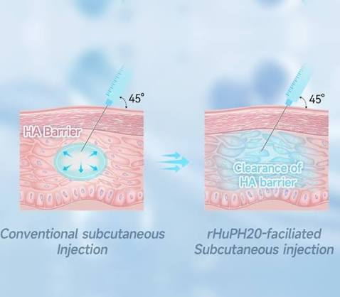
Figure 1. Conventional Subcutaneous Injection vs. rHuPH20-Enabled Subcutaneous Injection
Image description: The left panel shows a conventional subcutaneous injection, where the injected solution remains concentrated near the injection site because of the HA-rich extracellular matrix. The right panel illustrates rHuPH20 temporarily degrading local HA, allowing the injected solution to spread more efficiently through the subcutaneous tissue.
For drug delivery, however, this gel-like matrix can present a challenge. After a drug is injected into the subcutaneous tissue, it must move through this three-dimensional HA-rich network before reaching nearby blood and lymphatic vessels. Because the matrix restricts fluid movement, conventional subcutaneous injections are generally limited to relatively small volumes. Larger volumes can cause swelling, discomfort, or leakage from the injection site.
This physical limitation is one reason why many high-dose biologics have traditionally been administered intravenously.
"Molecular Scissors" Emerge
Understanding rHuPH20
rHuPH20 is derived from human PH20 hyaluronidase, an enzyme naturally associated with sperm cells. In human reproduction, PH20 participates in the degradation of HA-rich structures surrounding the egg, helping sperm penetrate the surrounding extracellular matrix.
Native PH20 is a membrane-associated protein. Its full-length precursor contains 509 amino acids and is anchored to the cell membrane through a glycosylphosphatidylinositol (GPI) anchor.
Researchers developed rHuPH20 by removing the signal peptide and C-terminal membrane-anchoring sequence while retaining the soluble, catalytically active region corresponding to amino acids 36-482 of the precursor. The resulting protein is a 447-amino-acid soluble enzyme that can be produced and formulated for pharmaceutical applications.
The recombinant enzyme was first described by Bookbinder and colleagues in 2006 in the Journal of Controlled Release.
Before recombinant human hyaluronidase became available, pharmaceutical applications relied in part on animal-derived hyaluronidases extracted from bovine or ovine tissues. These preparations could contain impurities such as immunoglobulins and proteases and were associated with a potential risk of hypersensitivity reactions.
Recombinant production in CHO cells provides a more controlled manufacturing process and enables production of a highly purified human enzyme, making rHuPH20 well suited for modern biologic drug-delivery applications.
Structure and Mechanism
How Does rHuPH20 Work?
Mature rHuPH20 contains 447 amino acids and has a molecular weight of approximately 61 kDa. It contains six potential N-glycosylation sites: N82, N166, N235, N254, N368, and N393.
These glycans are more than structural features. Research has shown that removing N-glycans can significantly reduce enzymatic activity, demonstrating that glycosylation plays an important role in rHuPH20 function.
This is also an important consideration when selecting an expression system. Mammalian cells such as CHO cells can provide the post-translational modifications required for proper protein folding and activity, whereas bacterial systems such as E. coli cannot reproduce mammalian glycosylation patterns.
In late 2024, researchers at the Korea Advanced Institute of Science and Technology reported the first cryo-EM structure of human PH20. The resulting structure was deposited in the Protein Data Bank as PDB 9JUB. The structure revealed a characteristic (β/α)₈-barrel catalytic domain along with an EGF-like domain, consistent with its classification in glycoside hydrolase family 56.
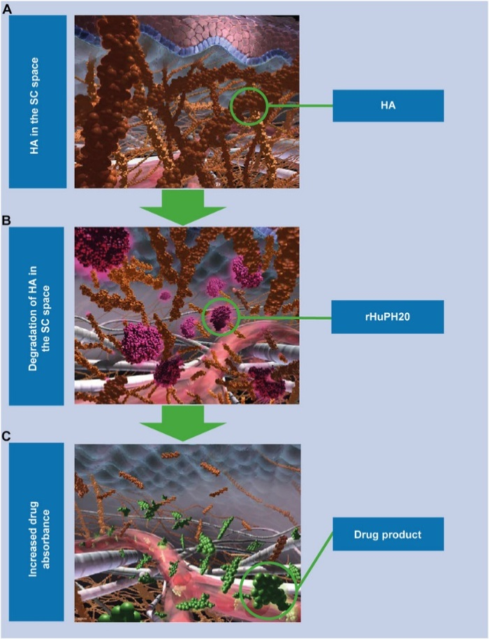
Figure 2. How rHuPH20 Enhances Subcutaneous Drug Delivery
Image description: (A) HA forms a dense extracellular matrix within the subcutaneous space. (B) After administration, rHuPH20 temporarily degrades the local HA network. (C) With the HA barrier temporarily reduced, therapeutic molecules can spread more efficiently through the tissue and enter systemic circulation through blood and lymphatic vessels.
Functionally, rHuPH20 is an endo-type hyaluronidase that hydrolyzes glycosidic bonds within HA. Rather than removing sugars sequentially from one end of the polymer, the enzyme cleaves within the HA chain, rapidly breaking the long polymer into smaller oligosaccharide fragments.
This process triggers a series of changes in the local tissue environment:
HA degradation → reduced matrix viscosity → improved fluid movement → enhanced drug dispersion → more efficient absorption
As the HA network is temporarily broken down, the extracellular matrix becomes less restrictive, allowing the injected drug to spread over a larger area and gain improved access to blood and lymphatic vessels.
The potential impact on drug delivery is significant. rHuPH20-enabled systems can accommodate substantially larger subcutaneous injection volumes than conventional approaches, with some applications supporting volumes ranging from tens to more than 100 mL.
From a pharmacokinetic perspective, improved subcutaneous dispersion can also affect the rate and extent of drug absorption, including parameters such as maximum plasma concentration and time to peak concentration.
In simple terms, rHuPH20 temporarily opens up the HA-rich extracellular matrix, creating more room for the therapeutic molecule to move.
Temporary, Local, and Reversible
Why Safety Matters
Breaking down an important component of the extracellular matrix may sound concerning. The key, however, is that rHuPH20-mediated HA degradation is localized and transient.
Preclinical studies have shown that rHuPH20 activity is concentrated near the injection site and declines rapidly following administration. HA itself also undergoes relatively rapid natural turnover, allowing the local extracellular matrix to recover within approximately 24–48 hours.
Immunogenicity is another important consideration. Because rHuPH20 is a recombinant human protein, its sequence is human-derived, although anti-drug antibody responses can still occur.
Studies have detected pre-existing antibodies capable of reacting with rHuPH20 in a subset of healthy individuals, and some treated patients also develop anti-rHuPH20 antibodies. To date, available clinical evidence has not established a clear clinical impact from these antibody responses. Safety reviews covering multiple studies have also supported the use of rHuPH20 as a subcutaneous drug-delivery technology.
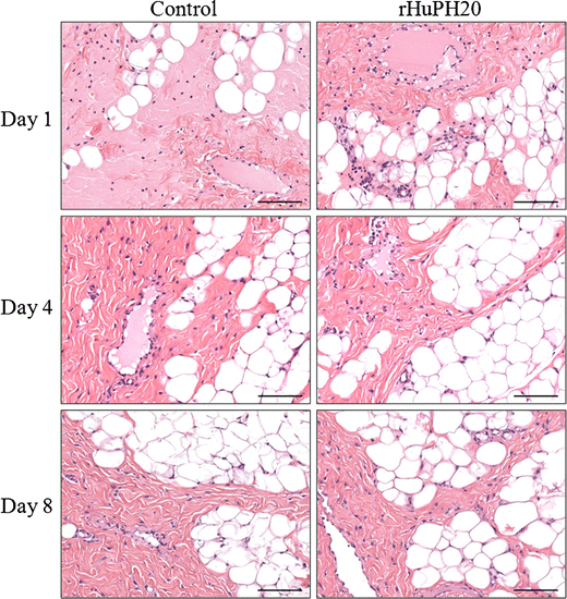
Figure 3. Histological Comparison of Injection Sites
Image description: H&E-stained sections compare subcutaneous tissue from control and rHuPH20-treated groups on Days 1, 4, and 8 following injection. The analysis evaluates whether rHuPH20 causes observable tissue damage or inflammatory changes and supports the characterization of its effects as local, temporary, and reversible.
How rHuPH20 Is Already Changing Drug Delivery
The value of rHuPH20 becomes especially clear when looking at its impact on treatment administration.
For example, the subcutaneous formulation of trastuzumab can reduce administration time from approximately 30–90 minutes to around 2–5 minutes.
For rituximab, subcutaneous administration can reduce treatment time to approximately 5–7 minutes. In clinical studies, a large proportion of patients reported a preference for subcutaneous administration, with reduced time spent in the clinic being one of the major reasons.
For daratumumab, the subcutaneous formulation can reduce an initial intravenous administration that may take several hours to an injection lasting only a few minutes. Clinical studies have also reported fewer administration-related reactions with the subcutaneous formulation.
Phesgo, a fixed-dose combination of pertuzumab and trastuzumab, is another example. Instead of receiving the two antibodies through separate intravenous infusions, patients can receive the combination subcutaneously, with the initial dose taking only several minutes to administer.
Meanwhile, HYQVIA uses recombinant human hyaluronidase to facilitate subcutaneous immunoglobulin replacement therapy. It can extend the interval between treatments and, after appropriate training, allow patients to administer treatment at home.
It is important to emphasize that rHuPH20 is not the therapeutic agent itself. It does not directly target tumors or replace immune components. Instead, it functions as a drug-delivery enhancer or permeation enhancer, helping the actual therapeutic molecule reach systemic circulation through a more convenient administration route.
In other words, rHuPH20 is the behind-the-scenes technology that helps make subcutaneous delivery possible for therapies that might otherwise require intravenous administration.
From Understanding an Enzyme to Engineering One
How Can Synbio Technologies Help?
Understanding how rHuPH20 works raises another question:
How do you go from a protein sequence or design concept to a functional, experimentally validated enzyme?
For proteins such as rHuPH20, the development process involves much more than simply synthesizing a gene and expressing the protein. A typical workflow may include:
Target and sequence selection → AI-assisted protein design → gene synthesis and codon optimization → expression vector construction → expression-system selection → protein expression → purification → structural and functional characterization
Each step can affect the final result.
A protein may be difficult to express. It may fold incorrectly, exhibit poor stability, have an inappropriate glycosylation profile, or show insufficient enzymatic activity. For proteins that depend on mammalian post-translational modifications, selecting the right expression platform is particularly important.
This is where Synbio Technologies brings together more than a decade of experience in gene synthesis and protein expression.
Our integrated workflow supports AI-assisted protein design, gene synthesis and codon optimization, expression vector construction, protein expression, purification, and structural and functional analysis.
For proteins requiring mammalian post-translational modifications, we offer CHO-based expression, as well as other expression platforms, including yeast, insect cells, and E. coli. This allows the expression strategy to be tailored to the biological characteristics of the target protein.
Whether you start with a protein sequence or a specific functional requirement, our team can support the downstream workflow, from vector construction and expression-system selection to protein production, purification, and functional validation.
The goal is simple: help shorten the path from protein concept to experimentally validated sample.
If your team is exploring hyaluronidases, delivery-enhancing enzymes, therapeutic proteins, or engineered enzymes that require precise glycosylation, high purity, or strong functional activity, Synbio Technologies can support your project across multiple stages of development.
Whether you are testing a new protein design, optimizing the activity or stability of an existing molecule, or developing a candidate for experimental validation, our gene synthesis, mammalian expression, protein engineering, and AI-assisted design capabilities can help turn molecular concepts into experimental data.
From sequence to protein. From design to validation. Synbio Technologies helps move your protein research forward.
Reference
1. Bookbinder LH, Hofer A, Haller MF, et al. A recombinant human enzyme for enhanced interstitial transport of therapeutics. J Control Release. 2006;114(2):230–241.
2. Im SB, Oh BH, et al. Cryo-EM Structure of Human Hyaluronidase PH-20. Proteins. 2025.
3. Hwang H, et al. Glycosylation of recombinant human hyaluronidase PH20 and its impact on enzymatic activity. [Journal information as indexed in PubMed].
4. Johnson & Johnson Medical Information. Recombinant human hyaluronidase PH20 (rHuPH20): Overview and mechanism of action.
5. Recombinant human hyaluronidase PH20: local and transient biodistribution following subcutaneous administration. PLOS ONE. 2021.
6. Recombinant human hyaluronidase PH20 for subcutaneous drug delivery: safety and clinical considerations. Expert Opin Drug Deliv. 2021.
7. Recombinant human hyaluronidase PH20: nonclinical safety assessment and effects of anti-rHuPH20 antibodies on fertility. J Control Release. 2024.
8. Clinical immunogenicity of recombinant human hyaluronidase PH20.
9. Anti-rHuPH20 antibodies in healthy individuals: baseline prevalence and characterization. BioDrugs.
10. Catalytic residues and mechanism of action of human hyaluronidases HYAL1 and PH20 at neutral pH. PLOS ONE. 2024.
11. Human PH20 hyaluronidase: hydrolysis mechanism, β-1,4 glycosidic bond cleavage, and oligosaccharide products. Front Bioeng Biotechnol. 2022.
12. BRENDA Enzyme Database. Hyaluronoglucosaminidase (EC 3.2.1.35).
13. Hyaluronic acid: biological functions and physiological roles. Frontiers.
14. U.S. Food and Drug Administration. HYLENEX (hyaluronidase recombinant) Prescribing Information. 2008.